Phang, I; Papadopoulos, MC
(2015)
Intraspinal Pressure Monitoring in a Patient with Spinal Cord Injury Reveals Different Intradural Compartments: Injured Spinal Cord Pressure Evaluation (ISCoPE) Study.
Neurocritical Care, 23 (3).
pp. 414-418.
ISSN 1556-0961
https://doi.org/10.1007/s12028-015-0153-6
SGUL Authors: Papadopoulos, Marios Phang, Isaac Sng Khai
|
Microsoft Word (.doc)
Accepted Version
Available under License ["licenses_description_publisher" not defined]. Download (53kB) |
||
![[img]](https://openaccess.sgul.ac.uk/108106/9.hassmallThumbnailVersion/Final.jpg)
|
Image (JPEG) (Supplementary Information)
Accepted Version
Available under License ["licenses_description_publisher" not defined]. Download (601kB) | Preview |
Abstract
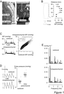
BACKGROUND: We recently described a technique for monitoring intraspinal pressure (ISP) after traumatic spinal cord injury (TSCI). This is analogous to intracranial pressure monitoring after brain injury. We showed that, after severe TSCI, ISP at the injury site is elevated as the swollen cord is compressed against the dura. METHODS: In a patient with complete thoracic TSCI, we sequentially monitored subdural ISP above the injury, at the injury site, and below the injury intraoperatively. Postoperatively, we simultaneously monitored subdural ISP and intraparenchymal ISP at the injury site and compared the two ISP signals as well as their Fast Fourier Transform spectra. RESULTS: Subdural ISP recorded from the injury site was higher than subdural ISP recorded from above or below the injury site by more than 10 mmHg. The subdural and intraparenchymal ISP signals recorded from the injury site had comparable amplitudes and Fast Fourier Transform spectra. Intraparenchymal pulse pressure was twofold larger than subdural pulse pressure. CONCLUSION: After severe TSCI, three intradural compartments form (space above injury, injury site, space below injury) with different ISPs. At the level of maximum spinal cord swelling (injury site), subdural ISP is comparable to intraparenchymal ISP.
Statistics
Actions (login required)
 |
Edit Item |


